
Facial ultrasound can help us find filler, assess a complication and guide treatment precisely. It is a diagnostic tool, not a treatment in itself. The value comes from understanding what the images show, how they relate to your symptoms and whether acting on them will help.
A complication consultation is the place to start. You can refer yourself or be referred by your practitioner. Dr MJ will assess your concerns and decide whether ultrasound would help.
Ultrasound uses sound waves to produce images beneath the skin. A probe is moved over the area with a little gel, allowing Dr MJ to examine the tissues in real time. It does not use ionising radiation.
A scan can help identify filler deposits and show their depth, distribution and relationship to surrounding structures. This is useful when filler is uneven, appears to have moved or remains after previous dissolving. Some materials have characteristic appearances that help distinguish filler types, although a scan cannot always establish the exact product or brand.
Ultrasound can help assess a lump, look for a collection of fluid and identify changes in the surrounding tissue. These findings can guide further investigation and management. They need to be interpreted alongside your treatment history and examination: an image alone cannot reliably tell us whether every lump is infected, inflammatory or simply a deposit of filler.
Doppler ultrasound can assess blood flow and help investigate vascular adverse events, where filler has affected the blood supply. Dr MJ accepts urgent complication referrals from patients and practitioners. If you suspect a vascular complication, contact our team immediately and make clear that the concern is urgent. Assessment and treatment should not be delayed while waiting for a routine scan appointment.
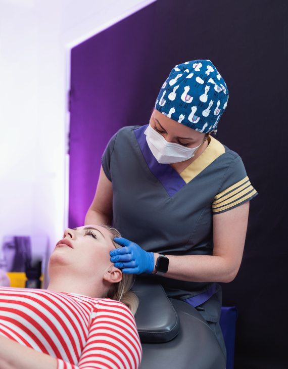
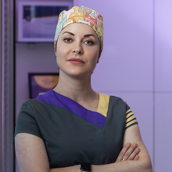
Dr MJ has treated thousands of complications and teaches facial ultrasound and guided treatment internationally. Patients and practitioners refer to her from across the UK, Europe and further afield. Her expertise is using ultrasound to understand complications and guide their management.
Our GE HealthCare Venue Go provides high-frequency imaging for detailed assessment of facial tissues. High-frequency probes can show fine detail that lower-frequency handheld ultrasound devices may miss. Image quality varies between devices and depends on the probe, settings and depth being examined.
The device is only part of the service. Using it to treat complications requires skilled interpretation and practical experience. Dr MJ combines both to decide what needs treating and perform targeted interventions where appropriate.
An ultrasound scan helps Dr MJ assess the problem and plan treatment. During ultrasound-guided treatment, she also uses the live image to watch the needle as she injects, helping her deliver the medication into the filler deposit.
For example, when dissolving hyaluronic acid filler, ultrasound can help guide hyaluronidase into an identifiable deposit. The enzyme breaks down the filler; the ultrasound helps direct its delivery. This can be particularly useful for a discrete lump, a deep deposit or residual filler after previous treatment.
Dr MJ is an expert in these guided interventions. They require the ability to interpret the image while controlling the probe and needle, together with a detailed understanding of facial anatomy and complication management.
Guidance is not needed for every injection. Some complications are better managed with treatment based on clinical examination, or with a combination of guided and unguided treatment. The decision follows your assessment.
Finding filler does not automatically mean it is causing your symptoms or needs removing. Ultrasound cannot explain every instance of swelling, prove that filler is obstructing lymphatic drainage or predict exactly how you will look after dissolving. It cannot make dissolve non-hyaluronic fillers, reverse ageing or repair every tissue change left by a longstanding complication.
Very small filler deposits may be below the level of detail ultrasound can show. Not seeing them does not mean they are absent: they may simply be too small to see. A scan that shows no identifiable filler therefore cannot guarantee that there is none left.
Ultrasound is a useful tool, but it cannot find or fix everything. It does not guarantee complete filler removal or a particular cosmetic result. Sometimes the most appropriate advice is to leave filler alone.
Our clinic is inclusive and welcomes anyone who needs help, without judgement. You can refer yourself or be referred by your practitioner. If you live far away, an online consultation gives you the chance to be heard and discuss your concerns before travelling. Ultrasound and guided procedures require an in-person appointment; your consultation will help establish what is appropriate.
Need help choosing an appointment? Call 0151 236 5166 or email reception@drmj.com. Follow @drmjclinic
Our team has helped many patients like you and will guide you towards the most appropriate booking. Assessment and treatment are separate elements of care; the proposed management and fees will be explained before you proceed.